Clinical and Procedural Effects of Transitioning to Contact Force Guided Ablation for Atrial Fibrillation

Daniel R. Frisch1, Sean J. Dikdan1

1 Thomas Jefferson University Hospital, Division of Cardiology, 111 S 11th St, Philadelphia, PA 19107.

Abstract

Background

A major innovation in atrial fibrillation (AF) ablation has been the introduction of contact force (CF) sensing catheters.

Objective

To evaluate procedural and clinical effects of transitioning to CF-guided AF ablation.

Methods

Consecutive AF ablation patients were studiedduring the period of time of transitioning from a non-CF to CF sensing catheter. Procedural data recorded was total radiofrequency time, time to isolate the left pulmonary veins (LPVs), and time to isolate the right pulmonary veins (RPVs). Clinically, the 3 and 12-month maintenance of sinus rhythm was noted and compared by: paroxysmal vs. persistent AF; CT scan LA volume more or less than 150 cc; CHA2DS2VASC more or less than 2; and LVEF more or less than 55%. Safety data was recorded as well.

Results

Total ablation times were shorter (113 vs.146 min, p=0.011)when using the CF catheters compared to non-CF ablations. This was driven by a decrease in both LPV (46 vs.72 min, p<0.001) and RPV time (54 vs. 75 min, p=0.002).The use of CF catheter did not change the overall percentage of patients in sinus rhythm at 3 and 12-months of follow up. However, sinus rhythm was more frequent at 12 months with CF ablation inpatients with an LA volume of more than 150 cc when compared to non-CF ablation (84.6% and 52.4%, p=0.03). There was no difference in outcomes with stratification by CHA2DS2VASC score or LVEF. No significant difference in complications was noted.

Conclusions

For AF ablation, the initial use of CF-sensing technology reduced procedure times with similar overall sinus rhythm maintenance at 3 and 12 months. CF improved 12-month outcomes in patients with an enlarged LA.

Key Words : Atrial Fibrillation, Ablation, Contact Force.

Correspondence to: Daniel R. Frisch, 925 Chestnut Street Mezzanine Philadelphia, PA 19107

Introduction

Electrical pulmonary vein isolation (PVI) for atrial fibrillation (AF) is an established and effective therapy.[1] Clinical trials have demonstrated that an ablation strategy is generally superior to antiarrhythmic medications for the treatment of AF.[2] Radiofrequency energy is the most common energy source used for ablation and is often delivered in a point-by-point fashion around the pulmonary veins. The original, non-irrigated catheters, recorded the temperature via a thermistor at the tip of the catheter and measuredimpedance changes over the duration of ablation. Later, irrigated catheters were introduced for improved ablation efficacy and safety.[3] A limitation of irrigated catheters is the inability to measure the temperature at the tissue level of ablation due to the intentional cooling effect of the irrigant on the catheter’s thermistor. The next major innovation in ablation technology, which was approved by the FDA in 2014,was the ability to measure catheter-tissue contact force (CF) in real-time and to use that information to guide ablation. BiosenseWebseter’s Smarttouch catheter was approved on February 25, 2014 and St. Jude Medical’s TactiCath was approved on October 27, 2014.[4,5] The use of contact force-guided ablation has been demonstrated to reduce ablation gaps and improve ablation effectiveness.[6,7,8]

Once the CF catheterswere approved at our institution we adopted them into use for pulmonary vein isolation in place of the irrigated, non-CF ablation catheters used previously. As with any new technology, there was a requisite period of introduction and transition. The purpose of this observational study was to assess the impact of the single variable of incorporating CF technology on procedural and clinical characteristics at the time of transition to this technology. The hypothesis was that the introduction of CF technology would improve both procedural and clinical aspects of PVI. We expected that cases would take less time, require less ablation, have fewer complications, and have better clinical outcomes with CF technology. The other aspects of ablation, including the ablation strategy, the personnel (a single attending electrophysiologist working with one of three fellows depending on the academic year), the other recording catheters, and the workflow remained the same.

Methods

This retrospective review included the period of time from July 2013 through November 2017, which was the timeframe for collection and follow up of 112 paroxysmal and persistent atrial fibrillation patients referred for ablation. Patients eligible for this study included consecutive patients who had undergone their first AF ablation with CF catheters and the consecutive group of patients who underwent their first AF ablation before CF catheters were available.Exclusion criteria included patients who underwent ablation for arrhythmias other than AF or who presented for a repeat procedure.

Procedural and clinical characteristics were collected from our institution’s electronic health record.Of the initial 112 patients designated for inclusion in the study, 51 patients underwent non-CF ablation and 61 underwent CF ablation; 7 patients in the non-CF group and 10 in the CF group were excluded from analysis because AF ablation was not performed or the presentation was for a repeat ablation procedure.The data were collected and stored securely in a password-protected database. The study was approved by our institutional review board.

Catheter Ablation Procedure

All patients were referred for catheter ablation of AF and provided written informed consent in accordance with institutional policy. Antiarrhythmic medications other than amiodarone were stopped three days before the procedure. In brief, femoral venous access was obtained and a multipolar catheter was placed in the coronary sinus and a diagnostic intracardiac ultrasound catheter (5.5 to 10 MHz; AcuNav; Biosense Webster, Diamond Bar, California) was placed in the right atrium. Two atrial transseptal punctures were performed, and an ablation catheter and a circular mapping catheter (Spiral; St. Jude Medical) were advanced into the left atrium. Three-dimensional electroanatomic mapping was performed using the Velocity system (St. Jude Medical).

All pulmonary veins were routinely isolated, typically as a pair. Ablation was performed in the carina between ipsilateral veins if isolation could not be achieved with wide area encirclement. Radiofrequency ablation was delivered with a 3.5-mm open-irrigated tip catheter or a 3.5-mm open-irrigated CF sensing catheter (TactiCath; St. Jude Medical, St.Paul, MN). For LA volumes exceeding 150 cc by cardiac CT a TactiCath 75 was used, and for a volume less than 150 cc a TactiCath 65 was used[9].With the non-CF catheter, radiofrequency was routinely delivered to lesions for 30 to 60 secondsto achieve a decrease in impedance of at least 5 to 10 Ohms at the ablation site. With the CF sensing catheter, ablation was performed with a flow of 17 cc/minute, power 20-25 watts, a goal of 10-40 g per lesion, and a goal of 400-500 g seconds per site (typically a lesion size index 4.5-5.5). Successful PVI was defined by the loss of all pulmonary vein potentials (entrance block) andfailure to capture the left atrium when pacing from sequential bipoles of the circular mapping catheter placed at the ostium of each pulmonary vein (exit block).Attempts at reinduction with burst pacing were performedand recorded.

The rationale for the use of CF catheters and the working parameters that we chose were determined by a number of published investigations. The first was the 2012 TOCCATA study, which was primarily a safety study for right and left atrial ablation using the same CF ablation catheter used in our study.[10] Investigators identified a force >100 g as a risk for perforation, which occurred in one patient. The EFFICAS I trial (2013) was designed to assess CF (using the TactiCath ablation catheter) and the ability to predict ablation gaps during ablation for AF.[11] The operators were blinded to the contact force data. The results established that a minimum CF (<10 g) and minimum force-time integral (FTI; <400 gs) were predictors of gaps in the ablation lesion set. To achieve durable lesions and to obtain a successful PVI, a target CF of 20 g was recommended, with an absolute minimum CF of 10 g and an absolute minimum FTI of 400 gs per individual ablation lesion. The SMART AF trial (2014) was designed for safety and effectiveness of the SmartTouch catheter.[6] In this trial, when the CF was between “investigator selected working ranges” >80% of the time, outcomes were 4.25 times more likely to be successful. In 2015, the EFFICAS II, which was designed based on the findings in EFFICAS I with unblinded operators using TactiCath, found that a CF of 20g and a minimum FTI of 400 gs reduced ablation gaps. The investigators found that fewer lesions were required, and lower fluoroscopy times were achieved with these parameters.[12] Finally, the TOCCASTAR study (2015) randomizedCF vs. non-CF for paroxysmal AF and looked at 1 year AF freedom after ablation (n=300) using TactiCath[7]. The authors noted that when optimal CF was used (≥90% of the lesions with a CF ≥10 g) outcomes were better (76% v. 58%) and fluoroscopy and ablation times were less. Support for the use of ablation catheters with CF parameters are supported by national guidelines.[13]

Follow up

Patients in this practice tend to remain within the health system. These patients were followed up periodically with routine office visits at up 1, 3, 6, and 12 months and both in between visits and beyond 12 months if there was a reportof symptoms. Standard electrocardiography was performedat each follow-up visit to assess AF status. Mobile cardiac outpatient telemetry monitors were used if indicated clinically. Phone calls and emails were encouraged with any symptoms.At 12-month follow up, data was able to be collected on 30 patients in the non-CF group and 38 patients in the CF group.

Study Endpoints

The primary procedural endpoints were total radiofrequency time, time to complete isolation of the left pulmonary veins, time to complete isolation of the right pulmonary veins, and inducibility to AF, atrial flutter, or other arrhythmias. The primary clinical endpoints were the presence of AF during the first 3 and first 12 months. Recurrence of AF was defined as 30 seconds or more of symptomatic or asymptomatic AF after ablation regardless of the pre-procedural burden or the patient’s perception of improvement after the procedure.

Statistical analysis

Statistical analysis of nominal variables was performed using t-tests, except in non-normal distribution where the nonparametric Wilcoxon rank-sum test was used. Analysis of categorical variables was performed with chi-square for overall comparison,adjusted residuals of the chi-square for multigroup comparisons, and Fisher’s exact testfor subgroup analysis given the cells of decreased number. Odds ratios and confidence intervals were also generated. All comparisons were tested at the level of α=0.05 except where indicated.For multigroup comparisons, the Bonferroni correction was applied, and used a level of α=0.01. Statistics were performed using SPSS 22.0 (Armonk, NY: IBM Corp.).

Results

Baselinecharacteristicsof the 95 included patients did not show any significant differences [Table 1]. The sample was predominantly men around the age of 60. Persistent AF comprised a larger proportion of the sample (60%)than paroxysmal AF.

Table 1. Patient demographics and baseline data
Contact Force (N=51) Non-Contact Force (N=44) p-value
Age, years mean (STD) 60.7 (9.8) 60.3 (8.8) 0.60
Male gender, no. (%) 40 (78.4) 29 (65.9) 0.13
Paroxysmal AF, no. (%) 21 (41.2) 17 (38.6) 0.48
CHA2DS2-VASc score median (IQR) 1 (1-2) 2 (1-3) 0.24*
Anti-arrhythmic drug use, no. (%) 16 (31.4) 10 (22.7) 0.24
Anticoagulation use, no. (%) 24 (47.1) 23 (52.3) 0.38
3D LA volume, mL 162.3 (39.9) 165.6 (46.3) 0.26
Left Ventricular Ejection Fraction, % 56.6 (13.3) 55.0 (15.8) 0.29

The nonparametric Wilcoxon rank-sum test was used to analyze CHA2DS2-VASc score, as medians instead of means were being compare.

Procedural Results

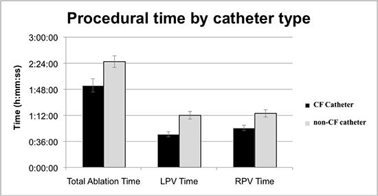
For the procedural analysis, data was complete for 86 patients. In each of the categories measured, there was a reduction in procedural time and total radiofrequency application time when a CF catheter was used [Table 2]. Theuse of a CF catheter significantly reduced the mean total ablation time by about 33 minutes (1 hour and 53 minutes compared to 2 hours and 26 minutes, p=0.011). LPV and RPV times were both significantlyshorter in the CF ablation group as well[Figure 1]. There was no difference in the ability to reinducesustained atrial fibrillation, non-sustained atrial fibrillation, or other arrhythmias between catheter types [Table 3].

Table 2. Procedural Times. Independent two samples two-tailed t-test of left, right, and total ablation time by catheter used.
Total Ablation Time LPV Time RPV Time
Contact Force Non-Contact Force Contact Force Non-Contact Force Contact Force Non-Contact Force
N 47 39 47 40 47 39
Mean 1:53:24 2:26:16 0:45:51 1:12:07 0:54:41 1:14:43
STD 1:03:38 0:51:11 0:24:44 0:33:55 0:25:28 0:31:33
p-value 0.011 <0.001 0.002


Table 3. Arrhythmia Inducibitily. Independent two samples two-tailed t-test of left, right, and total ablation time by catheter used.
Contact Force (N=46) Non-Contact Force (N=37) Chi-squared P value
Non-inducible 21 13 0.094 .30
AF 6 8 .33
Non-sustained AF 2 3 .47
Typical AFL 17 9 .22
Atypical AFL 0 4 .02

* Inducibility testing was not performed on 1 patient in the Contact Force group and 3 patients in the non-Contact Force group. These are excluded. * Bonferroni correction method was used to correct for multiple comparison post-hoc (adjust p-value=0.05/5= 0.01). No significant differences were found.

Figure 1. Graph of total, left pulmonary vein, and right pulmonary vein ablation time by catheter used.


Clinical Results

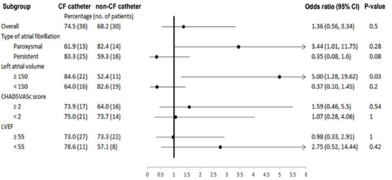
We chose 2 time points to evaluate for AF recurrence: 3 months—frequently considered the blanking period—and 12 months after ablation. No differencein the percentage of patients in sinus rhythm was detected between the CF and non-CF groups (74.5% and 68.2%, respectively; p=0.50) at the 3-month follow up period [Table 4a].The overall 12-month incidence of sinus rhythm was also not significantly different [Table 4b]; [Figure 2]).Subgroup analysis done at 12-month follow up showed that sinus rhythm was more frequent with CF compared to non-CF in patients with an LA volume greater than 150 cc compared (84.6% and 52.4%, respectively; p=0.03). There was no difference in outcomes with stratification by CHA2DS2VASC score or LVEF.

Table 4a. Clinical Outcomes. Patients in sinus rhythm after 3-months based on disease characteristics and catheter used.*
Contact Force Non-Contact Force P value
Overall patients in sinus rhythm, % (n) 78.4 (40) 68.2 (30) 0.26
Type of AF Paroxysmal 71.4 (15) 70.6 (12) 1.00
Persistent 83.3 (25) 66.7 (18) 0.22
LA volume, cc ≥ 150 76.9 (20) 61.9 (13) 0.34
< 150 80 (20) 73.9 (17) 0.74
CHA2DS2VaSc score ≥ 2 82.6 (19) 64.0 (16) 0.20
< 2 75.0 (21) 73.7 (14) 1.00
LVEF, % ≥ 55 78.4 (29) 80.0 (24) 1.00
< 55 78.6 (11) 42.9 (6) 0.12

* Chi-square used for overall analysis while fisher's exact test used for subgroup comparisons as some cells contained numbers <10

Table 4b. Clinical Outcomes. Patients in sinus rhythm after 12-months based on disease characteristics and catheter used.*
Contact Force Non-Contact Force P value
Overall patients in sinus rhythm, % (n) 74.5 (38) 68.2 (30) 0.50
Type of AF Paroxysmal 61.9 (13) 82.4 (14) 0.28
Persistent 83.3 (25) 59.3 (16) 0.08
LA volume, cc ≥ 150 84.6 (22) 52.4 (11) 0.03
< 150 64.0 (16) 82.6 (19) 0.20
CHA2DS2VaSc score ≥ 2 73.9 (17) 64.0 (16) 0.54
< 2 75.0 (21) 73.7 (14) 1.00
LVEF, % ≥ 55 73.0 (27) 73.3 (22) 1.00
< 55 78.6 (11) 57.1 (8) 0.42

*Chi-square used for overall analysis while fisher's exact test used for subgroup comparisons as some cells contained numbers <10

Figure 2. Forrest plotcomparing patients in sinus rhythm at 12 months based on catheter used and atrial fibrillation characteristics.


Safety Results

Overall, there was no observed increase in complications with the introduction of CF ablation. Pericardial effusion with or without the need for pericardiocentesis occurred in 3/45 = 7% of patients prior to the introduction of CF catheter and in 1/52 = 2% of the patients who underwent ablation with a CF catheter (p=0.24). There were no strokes, deaths, bleeding episodes requiring transfusion, esophageal injuries, or phrenic nerve injuries in either group.

Discussion

The intent of this study was to quantify the impact of the introduction of CF technology at the time of transition to this technology on procedural and clinical aspects of PVI. The ablation strategy, techniques, and workflow were the same but the catheter, specifically the ability to measure CF, was different. This strategy minimized confounding by other variables (e.g. changing the ablation strategy, the ablation modality, or the primary operator).

The main findings in this study of transitioning to the use of contact force catheters for atrial fibrillation ablation are that (1) procedure times, ablation times, and time to pulmonary vein isolation is reduced when contact force catheters are used; (2) clinical outcomes are similar, and perhaps improved in patients with large LA volumes; and that (3) complications rates were not increased.

As is the case with introduction of many new technologies, a learning curve is often required to become comfortable and demonstrate proficiency, thus maximizing the benefit of the innovation. Clearly, if new technology is difficult to use or if it is associated with complications it is unlikely to succeed. With the introduction of the contact force parameter there was a novel ablation parameter to follow. At times the tactile feel of the catheter would be discordant to the measured force. That is, the feeling of “heavy” force sometimes equated to a low force readings and vice versa. Confidence in the contact force recordings and calibration sometimes meant deciding which parameter (tactile feel or recorded force) represented the optimal ablation scenario. Because of this unfamiliar dilemma due to inexperience, we were reminded of the so-called “July phenomenon” (when there is a perceived decrease in the quality of health care at the start of the North American academic year for medical training).[14] Although the July phenomenon has been largely refuted,an abundance of evidence that “surgeon volume” matters across a range of operations including electrophysiology procedures.[15,16]

We found that, somewhat paradoxically, the initial use of a new ablation catheter improved procedural time and some effectiveness endpoints immediately, without the benefitof a large volume of cases. This reassured us about any concernswe had about slowingour workflow. Measuring contact force added usable information to the ablation strategy, and it also reduced ablation times, which ultimately improved workflow. The decreased procedural duration times were apparent almost immediately with adoption the contact force technology. Prior studies have shown a decrease in procedural time, but without differentiating between LPV and RPV times.[17,18]

Our study found that patients with large LA volumes (which we defined based on a previous investigation)undergoing AF ablation with a CF catheter were more likely to remain in sinus rhythm at 12 months.[9] This difference was driven by two CF patients converting to sinus between the 3 and 12 month period and two non-CF patients who convert from sinus back to AF in the same time period.One long term study has shown PV reconnection in both CF and non-CF ablated patients owing primarily to the RPV that negated a significant difference in atrial arrhythmia free survival[19].CF ablation has been shown to improve outcomes in patients with paroxysmal AF in large studies[20-22]and subsequent research on persistent AF has shown a benefit as well.[23] Patients with exclusively persistent AF and large LA enlargement, however,have been shown not to have an increase in sinus rhythm at 12 months.[24] Our analysis did not compare these two covariates directly, and it is possible that the advantage seen in our study was due to patients with large LA volumes and specifically paroxysmal AF.

Our complication rates were low in both arms, and similar to those of other studies involving CF ablations.[23,24]Larger studies with longer follow up have seen a reduction in complications with CF ablation so it is possible that we lacked significant enough power to detect a difference in complication rate[24].

Thisstudy has several limitations.First, the sample size is small. Despite initially selecting 112 charts, only 68 patients who met inclusion criteria completed their 12-month follow up. The patients lost to follow up appear in proportion between both the CF and non-CF group, but nevertheless this may result in unintended selection bias. Second, despite outpatient telemetry monitoring and regular electrocardiography it remains possible that patients had recurrences that asymptomatic and unrecorded. Third, subgroup analysis was not performed on procedural outcomes. Certain patient characteristics may have impacted procedural times. Finally, mean times to perform each AF ablation were reported. Changes in procedural time may have occurred towards the end of the CF group as the operator became more familiar with technology.

Conclusions

For atrial fibrillation ablation, introduction of CF-sensing technology reduced procedure times with similar overall sinus rhythm maintenance at 12 months. Notably, CF improved 12-month outcomes in patients with an enlarged left atrium.

References

  • [ PubMed ]
  • [ PubMed ]
  • [ PubMed ]
  • [ PubMed ]
  • [ PubMed ]
  • [ PubMed ]
  • [ PubMed ]
  • [ PubMed ]
  • [ PubMed ]
  • [ PubMed ]
  • [ PubMed ]
  • [ PubMed ]
  • [ PubMed ]
  • [ PubMed ]
  • [ PubMed ]
  • [ PubMed ]
  • [ PubMed ]
  • [ PubMed ]
  • [ PubMed ]
  • [ PubMed ]
  • [ PubMed ]
  • [ PubMed ]
  • [ PubMed ]
  • [ PubMed ]
  • [ PubMed ]