Hybrid Ablation of Ventricular Tachycardia: a Single-Centre Experience.

Mindy Vroomen 1, Bart Maesen 2, 1, Mark La Meir 3, Justin G.L Luermans 1, Kevin Vernooy 1, Jos G. Maessen 2, Harry J. Crijns1, Laurent Pison1

1 Department of Cardiology.2 Department of Cardiothoracic Surgery, Maastricht University Medical Center and Cardiovascular Research Institute Maastricht, Maastricht, the Netherlands .3 Department of Cardiac Surgery, UZ Brussel, Brussels, Belgium.

Abstract

Background

The long-term results of endocardial and percutaneous epicardial catheter ablation of ventricular tachycardia (VT) in patients with structural heart disease are disappointing. Arrhythmia recurrence after ablation and VTs with an epicardial substrate remain a clinical challenge. The purpose of this manuscript is to elaborate on feasibility and potential advantages of a surgical hybrid ablation (i.e., combined endocardial and surgical epicardial ablation) based on our initial experience consisting of five cases.

Methods

Endocardial electro-anatomical voltage and activation maps were created (Carto, Biosense Webster, California, USA), and endocardial radiofrequency (RF) applications were applied at exit sites, low voltage areas and isthmi. Next, after surgical access, epicardial voltage and activation maps were produced in combination with visual assessment of the epicardial substrate. Epicardial low voltage areas, isthmi and exit sites were identified and ablated using RF energy.

Results

After the procedure, VT was non-inducible in 80% of the cases (4/5, in one case no induction was performed). No peri-procedural complications occurred. After a mean follow-up of 18 months, one patient remained in sinus rhythm without, and 2 with use of antiarrhythmic drugs. One patient needed a redo procedure after 21 months, and in one patient the amiodarone dose was raised because of 2 sustained VTs. After this additional treatment, both kept sinus rhythm.

Conclusions

Hybrid VT ablation is a safe and effective patient tailored procedure that comprises the major advantage of combining direct anatomical visualization and enhanced catheter stability with high-density 3D mapping. As a consequence, this procedure should be considered as a valid treatment option in complex VT management.

Key Words : ventricular tachycardia, hybrid ablation, endocardial-epicardial ablation, endo-epicardial ablation, surgical ablation.

Correspondence to: TB. Maesen, Department of Cardiothoracic Surgery PO Box 5800, 6202 AZ, Maastricht, The Netherlands

Introduction

Ventricular tachycardia (VT) is an important cause of cardiac morbidity and mortality. In structurally diseased hearts, like coronary artery disease, the mechanism of VT is predominantly based on macro reentry due to scars in the myocardial tissue. [1,2] Achieving transmural ablation lesions in these scars remains challenging because they often consist of fibrotic strands branching out into the myocardial wall. Examples of non-ischemic VT causing diseases are, among others, arrhythmogenic right ventricular cardiomyopathy (ARVC) and Chagas disease.[3]

Implantable cardioverter-defibrillators (ICDs) are critical to prevent sudden cardiac death, whereas preventing recurrence of the arrhythmia is of utmost importance for patient well-being and long-term survival.[4,5] Endocardial catheter ablation has emerged as an important option in preventive VT treatment.[6] Epicardial ablation usually is performed only in cases where endocardial mapping fails to identify the site of the arrhythmia, or if the source of the VT is suspected to be located in the epicardium, as suggested by electrocardiographic characteristics of the VT, the underlying disease or imaging. In case of suspicion of epicardial involvement, first-line simultaneous endo-epicardial ablation might be considered.[7,8] Epicardial access and ablation can be performed percutaneously using a subxiphoid puncture [9], however, dry pericardial puncture has its known complications (e.g. cardiac perforation, vessel and nerve injury and pneumothorax).[10]

Combining an endocardial approach with epicardial surgical access during the same procedure, i.e. hybrid VT ablation, could overcome the mutual technical challenges and result in enhanced visualization and characterisation of the substrate and superior outcome. This manuscript aims to describe the feasibility and potential advantages of this hybrid procedure. Based on our single-centre experience, a series of five complex cases with frequently recurring monomorphic sustained VTs of different aetiologies is discussed.

Methods

The five procedures were carried out between November 2013 and February 2016 in the hybrid operating room (OR) of the Maastricht University Medical Center, Maastricht, The Netherlands. The interventions were performed under general anaesthesia with double-lumen endotracheal tube placement for selective single lung ventilation. In case 3 a single-lumen tube was placed. All patients were placed in supine position on the operating table, with arms next to the body. A His bundle catheter (St. Jude Medical, Minnesota, USA) was positioned through a 6 Fr sheath in the right femoral vein, and a ThermoCool irrigated tip contact force mapping and ablation catheter (Biosense Webster, California, USA) was advanced through an 8 Fr sheath in the right femoral vein or artery. All patients except one (case 3), then underwent heparinization to maintain an activated clotting time above 300 seconds. In case of sinus rhythm (SR), an endocardial electro-anatomical voltage map (cut-offs 0.5 – 1.5 millivolt (mV)) was created and subsequently VT was induced through programmed ventricular stimulation via the His catheter, with or without administration of isoprenaline. If VT was induced and hemodynamically tolerated, an activation map was constructed (Carto, Biosense Webster). Radiofrequency (RF) applications (maximum output 40Watt (W)) were then applied at the exit sites (i.e. sites of presystolic potentials during VT, or pacing sites with identical QRS morphology to the clinical VT), isthmi (i.e. conductive myocardial tissue delineated by nonconductive tissue, 0.5-1.5mV) and low voltage areas (i.e. nonconductive tissue, <0.5 mV). If no VT was inducible, or if the patient became hemodynamically unstable after VT induction, only substrate modification was performed: ablation of local abnormal ventricular activities (LAVA), i.e. low voltage and fractionated potentials annotated after the QRS complex.[11]

Next, in the first two cases, the left lung was deflated, followed by left anterolateral mini-thoracotomy without rib spreading using a soft tissue retractor [Figure 1]. The serratus anterior muscle and the intercostal muscles were cleaved, the pectoralis muscle was spared. The pericardium was opened ventrally of the phrenic nerve. In the third case, a re-sternotomy was performed. In the last two cases the access to the heart was provided by one-sided thoracoscopy using three access ports. An epicardial activation and voltage map (Carto, Biosense Webster; cut-offs 0.5 – 1.5mV) of the area of interest, as identified by endocardial mapping, were created using the endocardial catheter (Thermocool irrigated tip, Biosense Webster). Epicardial low voltage areas, isthmi and exit sites were identified. Using the same catheter (30-40W), epicardial applications were performed.

After endo- and epicardial ablation repeated VT induction (ventricular stimulation with or without administration of isoprenaline) was performed. If necessary, mapping or ablation was repeated.

Figure 1 Intra-operative Images


Left: Left-sided view of the chest with anterolateral thoracotomy. The endocardial radiofrequency catheter is placed on the epicardial surface. Right: Left-sided view of the chest with thoracoscopy using three access ports. Right upper: Left-sided thoracoscopic view of the heart. D = diaphragm, A = ablation catheter, P = left lung, F = forceps, LV = left ventricle.

Results

Baseline demographic and clinical characteristics

In the following section each case will be discussed separately. A summary of the five cases is depicted in [Table 1].

Case 1 : A 70-year-old man suffering from ischemic VTs with recurrent ICD shocks despite use of amiodarone, underwent 3 endocardial VT ablations. Using an activation map, an isthmus was located and targeted in the middle of the inferoposterior to septal area of the left ventricle (LV) (areas 3/4/5/6 according to Josephson[12,13], [Figure 2]). Substrate modification of the inferior LV was performed during the second ablation. Since this neither provided long-term freedom of VT, and because of suspicion for an epicardial origin on ECG (wide QRS, pseudo delta wave, [Figure 3A]), a percutaneous epicardial ablation was proposed. Access to the pericardium with a subxiphoideal puncture failed (dry tap), and per hospital protocol surgical access is not performed in the electrophysiology (EP) lab. Therefore, only an endocardial ablation was conducted. The exits of 2 induced VTs were mapped at areas 7 to 10 (apical low lateral/ inferoposterolateral/anterolateral/lateral basal), where several RF applications were performed. Another VT led to supplemental RF applications at areas 5/6/7 (inferior/inferoposterior/apical low lateral).

In the next days the patient again suffered from sustained VTs and therefore underwent a hybrid VT ablation in the OR, during which an endo- and epicardial ventricular substrate modification (LAVA) was performed (Fig.3B-C-D). A total of 55 applications were performed endocardially in area 5/6/7 (inferior/inferoposterior/apical low lateral), and 19 epicardially. The duration of each application was 30 seconds, the average power 36W and the maximum power 41W. No VT could be induced at the end. The patient was discharged after 3 days in the intensive care unit and 4 days on the regular ward.

After the hybrid procedure the incidence of VT while on beta-blockade was significantly reduced to sporadic sustained VT less than once per year. Twenty-one months after the ablation a sustained VT recurred (RBBB, undefined axis, R/S transition V4, CL440ms), leading to repeat endocardial ablation inferior/lateral (areas 5 to 10). In the following 18 months the patient did not suffer recurrences.

Figure 2 Endocardial Areas of Josephson[13]


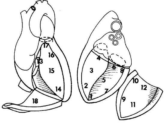
Left ventricle: 1 = apex, 2 = apical septum, 3 = mid septum, 4 = basal septum, 5 = inferior, 6 = inferoposterior, 7 = apical low lateral, 8 = inferoposterolateral, 9 = anterolateral, 10 = lateral basal, 11 = midanterior, 12 = superior basal. Right ventricle: 13 = tricuspid annulus/basal septum, 14 = apex, 15 = mid septum, 16 = anterior septum, 17 = outflow tract, 18 = lateral/free wall.
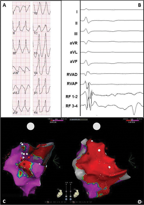
Figure 3 Electrocardiogram and electro-anatomical maps of case 1


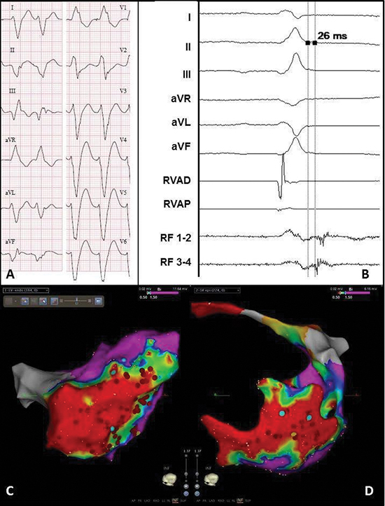
A. Twelve-lead electrocardiogram showing a ventricular tachycardia (115bpm) with right bundle branch block morphology, right axis and early R-wave transition, suggesting a lateral mid-apical origin in the left ventricle. Pseudo delta wave and wide QRS suggest epicardial origin.

B. Recording obtained in sinus rhythm during the procedure showing local abnormal ventricular activation at the RF catheter. RVAD/P = right ventricular apex distal/proximal, RF = radiofrequency.

C. Inferior view of the bipolar endocardial voltage map showing a low voltage area from inferoposterior to low apical in the lateral wall (area 5/6/7). Red dots = RF applications.

D. Inferior view of the bipolar epicardial voltage map showing a low voltage area consistent with the endocardial map. Red dots = RF applications.

Case 2 : An 80-year-old male with a history of MI and bilateral pulmonary vein isolation for paroxysmal AF, presented with a sustained VT two years after cardiac surgery. Despite amiodarone, recurrences with a ventricular rate below the detection zone of the ICD recurred. The LV voltage map during the first endocardial VT ablation showed a low voltage area from area 5/6 (inferior/inferoposterior) to 7/8 (apical low lateral/inferoposterolateral), and an early activation at area 7 (apical low lateral). RF applications in that area terminated the VT. Substrate modification in the exit zone of the VT was also performed (ablation line around the border zone and additional applications at areas 7/8, apical low lateral/inferoposterolateral). Ten days later the same VT recurred, followed by endocardial substrate modification in area 9 (anterolateral).

One month later a hybrid VT ablation was performed for recurrences despite the use of amiodarone and metoprolol ([Figure 4A]). Endocardially, the exit site of the clinical VT was mapped at area 8 (inferoposterolateral). A second VT with a more septal exit could be induced as well. The exit zones and late potentials [Figure 4B] were ablated (14 applications, mean 70 seconds, average output 39W). Adhesions and grafts of the previous cardiac operation, hindered the procedure, but safe mapping and ablation could be performed. The epicardial voltage map correlated with the endocardial map [Figure 4C-Figure 4D]. The exit was also mapped at area 8 (inferoposterolateral), and this zone and late potentials were ablated epicardially (11 applications, mean 45 seconds, average output 34W). Because of transient ST-segment elevations inferior, no more VT induction or ablation was performed. After 2 days in the intensive care unit, and 2 days on the regular ward, the patient was discharged.

During 12 months follow-up the patient did not suffer from any recurrences, while using metoprolol 50mg and amiodarone 100mg once per day.

Figure 4 Electrocardiogram and electro-anatomical maps of case 2


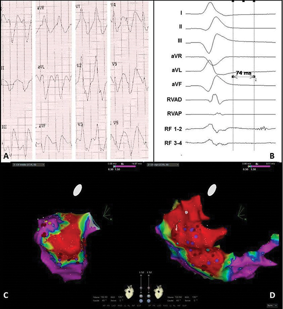
A. Twelve-lead electrocardiogram showing a ventricular tachycardia (150bpm) with right bundle branch block morphology, right axis and normal R-wave transition suggesting a lateral-basal origin in the left ventricle. Pseudo delta wave and wide QRS suggest epicardial origin.

B. Recording obtained in sinus rhythm during the procedure showing local abnormal ventricular activation. RVAD/P = right ventricular apex distal/proximal, RF = radiofrequency.

C. Inferior view of the bipolar endocardial voltage map showing low voltages inferior/inferoposterior (area 5/6). Red dots = RF applications, purple dots = late potentials.

D. RAO view of the bipolar epicardial voltage map showing low voltage area practically consistent with the endocardial map, though more extensive at the lateral wall (area 7/8). Red dots = RF applications, purple dots = late potentials.

Case 3 : A 56-year-old man was admitted with a therapy refractory incessant VT. On echocardiogram LV function was decreased to 30%, and the right ventricle (RV) was minimally dilated with also decreased function. ARVC was not seen on magnetic resonance imaging (MRI), but 2 of the major ARVC criteria were present: biopsy showed minimal fibrosis and myocytes with vacuolisation, and ECG showed sustained VTs of LBBB morphology with superior axis and inverted T waves in the right precordial leads in SR. Due to hemodynamic instability the patient was connected to extracorporeal life support. An endocardial electro-anatomical map of the RV (Ensite NaVX, St. Jude Medical) showed focal activation (i.e. earliest activation time -20mS during VT) at areas 15 (mid-septum) and 18 (lateral), and RF applications at and between those areas terminated the VT.

When trying to reduce the flow of the external support in the next days, VTs of morphology 1 and 2 recurred. These could not be induced during the second and third endocardial ablation, and a RV bipolar voltage map (Carto, Biosense Webster) showed no zones of low voltages. However, using unipolar signals an extensive low voltage zone (<5.5mV [14]) at areas 17 (outflow tract) and 18 (lateral) was seen. Epicardial access failed because the injected contrast could not be seen in the epicardium. Therefore only endocardial ablation was performed at the lateral area 18 (48C, 40W). The last application resulted in a steam pop, substantial loss of blood via the inserted pigtail in the pericardium, and hemodynamic instability. A hole in the RV free wall was sutured via a sternotomy performed in the EP lab. One day after discontinuation of external support, a therapy refractory sustained VT recurred (morphology no. 3). Since the VT terminated on arrival in the EP lab and could not be induced, it was decided to perform a hybrid VT ablation via resternotomy in the OR. After administration of isoprenaline a sustained VT was induced [Figure 5A-Figure 5B]. The endocardial activation map showed prepotentials at areas 13, 14 and 18 (basal, apical and RV free wall, [Figure 5C]), but hereafter the VT stopped. The bipolar epicardial voltage map showed low voltages in basically all areas of the RV ([Figure 5D]). Since the RF catheter could not be stabilized well on the beating hart, it was changed for a cryo-pen (AtriCure, Ohio, USA). A lesion of 4x4cm was made (8 applications of 120ms) at area 18 (lateral). No additional endocardial ablation was performed because of the recent perforation. No VT could be induced at the end of the procedure. After 6 days in the intensive care unit, the patient was transferred to his own hospital. Before discharge an ICD was implanted.

During one-year follow-up the patient maintained SR using metoprolol 100mg once per day.

Figure 5 Electrocardiogram and electro-anatomical maps of case 3


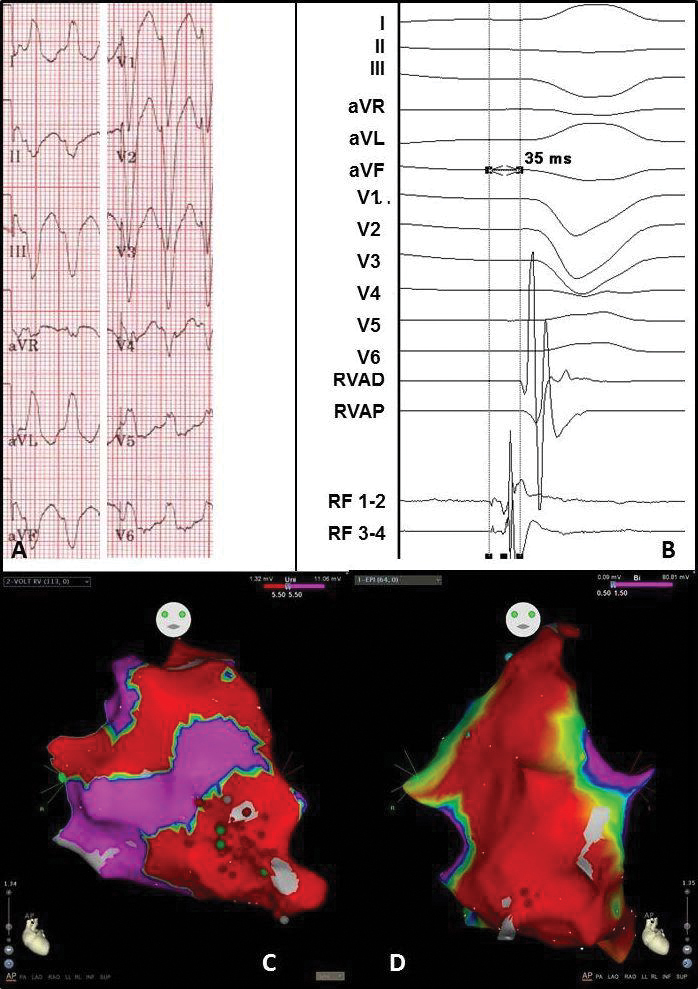
A. Twelve-lead electrocardiogram showing a ventricular tachycardia (180bpm) with left bundle branch block morphology and superior axis, suggesting origin in the right ventricle (RV) free wall.

B. Recording obtained in sinus rhythm during the procedure showing prepotentials. RVAD/P = right ventricular apex distal/proximal, RF = radiofrequency.

C. Anterior-posterior (AP) view of the unipolar endocardial voltage map showing a low voltage area at the apex and RV free wall (area 14 and 18). Red dots = RF applications

D. Anterior-posterior (AP) view of the bipolar epicardial voltage map showing a low voltage area at basically the total apex and RV free wall (area 14 and 18). Red dots = RF applications

Case 4 : A 67-year-old man, recently diagnosed with paroxysmal AF, was admitted to the hospital suffering a symptomatic monomorphic VT causing hemodynamic instability. Magnetic resonance imaging showed hypokinesia and subepicardial fibrosis of the basal inferolateral and inferior walls, and a normal RV [Figure 6]. DNA analysis showed a mutation in the PRKAG2 gene (c432dup mutation). Most probably this relates to a pathogenic mutation, however this mutation has not been described before nor is it known in genetic databases. A few months after ICD implantation, recurrent VTs (same morphology) occurred. Since there was clear evidence for an epicardial origin (imaging and ECG), it was decided to perform a left-sided thoracoscopic hybrid ablation. Macroscopically only minimal epicardial fibrosis was visible at area 8 and 10 (inferoposterolateral and lateral basal). The endocardial unipolar voltage map showed low voltages (<8.3mV, [15]) at those areas ([Figure 7C]), and the epicardial unipolar map also at the anterolateral area 9 ([Figure 7D]). No VT could be induced. Eight epicardial RF applications were performed at areas 9/10 (anterolateral/lateral basal) with a mean of 35seconds per application (average output 30W). Due to the proximity of a coronary artery, area 8/9 (inferoposterolateral/anterolateral) could not be ablated epicardially and was only ablated endocardially (6 applications with a mean of 60seconds and an average output of 25W). Because of AF during the procedure, isolation of the right and left pulmonary veins in pairs (bipolar clamp, AtriCure) was performed via the left-sided thoracoscopy. No VT could be induced at the end of the procedure. After 1 days in the intensive care unit, and 6 days on the regular ward, the patient was discharged.

In the first three months after ablation two sustained VTs recurred (RBBB, inferior axis, absent R/S transition, CL 340 ms). Blood analysis showed that the amiodarone level was subtherapeutic, and for one week the dose was raised to 600mg daily. The following year he maintained SR.

Figure 6 Cardiac magnetic resonance image of case 4, short axis.


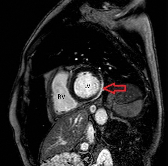
RV = Right Ventricle. LV = Left Ventricle. Red arrow: area of fibrosis (white).
Figure 7 Electrocardiogram and electro-anatomical maps of case 4


A. Twelve-lead electrocardiogram showing a ventricular tachycardia (206bpm) with right bundle branch block morphology, right inferior axis, and positive QRSs in all precordial leads, suggesting origin in the basal lateral wall of the left ventricle. Pseudo delta wave and wide QRS suggest epicardial origin.

B. Epicardial recording obtained in sinus rhythm during the procedure showing local abnormal ventricular activation. RVAD/P = right ventricular apex distal/proximal, RF = radiofrequency.

C. Posterior-anterior (PA) view of the unipolar endocardial voltage map showing a low voltage area inferoposterolateral and lateral basal (area 8/10). Red dots = RF applications.

D. PA view of the unipolar epicardial voltage map showing more extensive low voltages at the lateral basal area 10. Red dots = RF applications.

Case 5 : An 18-year-old male with working diagnosis ARVC (pre-syncopes, minimal dilated RV and frequent premature ventricular complexes (PVCs)), had recurrent ICD shocks and showed many non-sustained VTs. Despite the patient was suspected for being noncompliant to antiarrhythmic therapy and for excessive use of cannabis and alcohol, an endocardial VT ablation was performed. The bipolar voltage map showed low voltages at the basal septum, outflow tract and basal inferior RV (areas 13/17/18), wherefore ventricular substrate modification. No VT could be induced afterwards. In the following period the amount of runs of PVCs, non-sustained VTs and VTs increased ([Figure 8A-Figure 8B]), and a hybrid VT ablation via right-sided thoracoscopy was performed. No VT could be induced, but the bipolar endocardial voltage map showed low voltages at areas 13/15-18 (basal septum/mid septum/anterior septum/outflow tract/basal inferior,[Figure 8C]). The bipolar epicardial voltage map showed low voltage zones and LAVA at area 13 (basal septum), and low voltages at area 17 (outflow tract, Fig 8D). The unipolar endocardial map showed low voltages (<8.0mV) involving almost all areas. A first PVC was endocardially mapped and epicardialy and endocardially ablated at area 18 using RF (7 applications with a mean of 51 seconds per application, an average power of 34W and maximum power of 38W). A second PVC could be mapped epicardially at area 17, and was ablated epicardially using RF (5 applications with a mean of 53 seconds, average power of 71W and maximum power of 75W). No VT could be induced at the end of the procedure. After 3 days on the regular ward the patient was discharged.

During 12 months follow-up SR was maintained using sotalol 40mg twice per day.

Figure 8 Electrocardiogram and electro-anatomical maps of case 5


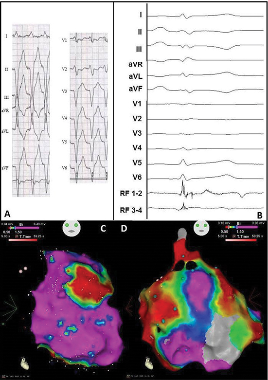
A. Twelve-lead electrocardiogram showing a ventricular tachycardia (110bpm) with left bundle branch morphology, superior axis and R/S transition in V3, suggesting origin in mid RV. No specific signs for epicardial origin.

B. Epicardial recording obtained in sinus rhythm during the procedure showing local abnormal ventricular activation. RVAD/P = right ventricular apex distal/proximal, RF = radiofrequency.

C. Anterior-posterior (AP) view of the bipolar endocardial voltage map showing a low voltage area from the basal inferior RV to mid inferior RV and RV outflow tract (areas 13/15-18). Red dots = RF applications.

D. AP view of the bipolar epicardial voltage map showing a more extensive low voltage area consistent with the endocardial map. Red dots = RF applications.

Table 1. Patient and procedural characteristics
Pre-operative Operative Post-operative
Case Gender Age VT Etiology Location EF (%) First VT Other VT AAD ICD CA Access Scar FU
1 M 70 Ischemic RCA, LCX, LMA 37 LBBB, superior axis, R/S V2, CL 320ms 2/3. RBBB, inferior axis, CL 310-360ms, 4. LBBB superior axis, R/S V6, CL 390ms, 5. LBBB, superior axis, R/S V2, CL 530ms Amiodarone + 3 Left Lateral AL/I 1 CA, AAD-
2 M 80 Ischemic LAD, LMA, RDP 41 RBBB, inferior axis, R/S V4, CL 400ms 2. superior axis, CL 395ms, rest unknown Amiodarone + 2 Left Lateral IL SR, AAD+
3 M 56 ARVC N.A. 35 LBBB, superior axis, R/S V5, CL 560ms 2. LBBB, superior axis R/S V4, CL 440ms 3.LBBB, inferior axis, R-S V4, CL 420ms None - 3 Sternotomy N.A. SR, AAD-
4 M 68 Unknown N.A. 57 RBBB, inferior axis, R/S V5, CL 230ms - None + 0 Left thoracoscopy N.A. VT, AAD+
5 M 35 ARVC N.A. 57 LBBB, inferior axis, R/S V3, CL 380ms 2. PVC: LBBB, superior axis, R/S V5-6 3. PVC: LBBB, inferior axis, R/S V5-6 Sotalol + 1 Right thoracoscopy N.A. SR, AAD+

EF = ejection fraction, VT = ventricular tachycardia, AAD = antiarrhythmic drug, ICD= implantable cardioverter-defibrillator, CA = catheter ablation, FU = follow-up, M = male, ARVC = arrhythmogenic right ventricular cardiomyopathy, RCA = right coronary artery, LCX = left circumflex artery, LMA = left marginal artery, LAD = left anterior descending artery, RPD = right posterior descending artery, N.A. = not applicable, LBBB = left bundle branch block, RBBB = right bundle branch block, R/S = r-wave/s-wave transition, CL = cycle length, AL = anterolateral, I = inferior, IL = inferolateral, SR = sinus rhythm

Discussion

In this report we describe five cases of patients with recurrent sustained VTs of variable etiology, treated with a hybrid VT ablation. Hybrid VT ablation is a novel technique as it combines endocardial and surgical epicardial ablation. In all patients, antiarrhythmic medication failed or was not tolerated, and four patients experienced arrhythmia recurrence after previous endocardial ablation. In two cases no percutaneous epicardial access could be obtained.

No perioperative complications occurred. ne patient underwent a redo endocardial ablation for recurrent sustained VT after 21 months. In another patient two sustained VTs recurred under a subtherapeutic amiodarone dose which was treated with a dose increase. One patient remained in sinus rhythm without, and two with the use of antiarrhythmic drugs.

Surgical and hybrid VT ablation

Since beating heart surgical and hybrid VT ablations are not often performed, studies reporting on these procedures are scarce. In a retrospective multicentre study among 913 VT ablations, Sacher et al. found that 17% of the procedures involved epicardial mapping and ablation.[1] In the vast majority (88%) of epicardial ablation, a percutaneous approach was preferred. A subxiphoidal access, mainly for catheter based mapping or ablation, was only used in 1.5% of the cases, while surgical treatment during concomitant cardiac surgery procedures was even lower (0.7%). Soejima et al. were the first to show that subxiphoidal surgical access provides successful entry for epicardial catheter-based mapping in patients with a failed percutaneous approach.[16] In a population of 444 treated VT patients, Sarkozy et al. reported this approach in 13 patients.[17] However, in only 6% of the total population they were able to successfully ablate the VT circuit epicardially, which is substantially less compared to endocardial circuits. Mathuria et al. reported a case of an epicardial ablation, under direct surgical vision of the epicardium, via a limited anterior thoracotomy by using an unidirectional epicardial bipolar RF device (Coolrail, AtriCure).[18] Also epicardial VT ablations, under direct surgical vision, using endocardial RF catheters combined with video-assisted mini-thoracotomy and left lateral thoracotomy approaches have been reported.[19,20] Furthermore, one case is described in which both endo- and epicardial mapping and ablation via sternotomy was performed surgically.[21] Michowitz et al. reported on a minimal invasive hybrid VT ablation under direct surgical vision in three patients via limited anterior thoracotomy.[22] Recently Li et al. compared the subxiphoidal and thoracotomy approach with the percutaneous approach, showing no significant difference in complications or outcome.[23] These publications show there are numerous options to perform VT ablation, giving sufficient possibilities for a patient tailored therapy.

Patient selection

It is important to determine which patients might benefit from an epicardial treatment since VT circuits that can be successfully ablated from the epicardium are less common than those that can be addressed from the endocardium. Also, endocardial circuits are seen in 50% of the patients in whom VT was ablated successfully from the epicardium, and additional ablation of the endocardium is often required.[17] Studying 19 patients, Tung et al. demonstrated that percutaneous epicardial in combination with endocardial ablation in patients with ischemic and non-ischemic cardiomyopathy was not superior to endocardial ablation alone after 12 months of follow-up.[24] On the contrary, Di Biase et al. showed in 92 patients that recurrence rates were lower in patients treated with endo- and epicardial ablation compared to endocardial ablation alone during a mean follow-up of 25 months (19% versus 47%).[25]

To date, epicardial ablation is mainly performed after previously failed endocardial ablation, in cases in which the epicardium seems to be the source of VT during endocardial ablation, or in which endocardial access is not possible. It can, however, also be considered to be performed as a first-line epicardial or hybrid VT ablation in patients with high suspicion of an epicardial circuit based on the underlying disease (like in Chagas disease, e.g.).[7,8,26,27] Sacher et al. observed the highest prevalence of an epicardial substrate in patients with a diagnosis of ARVC (41%), followed by non-ischemic dilated cardiomyopathies (35%) and ischemic heart disease (16%).[1] Suspicion of an epicardial origin could besides by the diagnosis, also be raised based on the ECG. As Berruezo et al. showed in a population consisting of 65% to 90% ischemic VTs, a VT originating from the epicardium produces a pseudo delta wave that corresponds to widening of the initial part of the QRS-complex.[28] The group of Valles et al. assessed ECG criteria for epicardial origin in a group of non-ischemic VTs and developed a 4-step algorithm.[29] Cardiac MRI can also be helpful in identifying patients with an epicardial substrate (like in case 4 of this report).[30]

Surgical approach

There are several reasons to consider a surgical approach for VT ablation. The most common is failure of a percutaneous approach, which is unsuccessful in 10% of the cases.[1] Another reason could be the anatomy, for example necessity to ablate close to the phrenic nerve or coronary arteries. In the latter case, a surgical approach with direct visualization of the epicardium, allows evaluating whether an epicardial ablation can be performed safely.[10] Here we describe a surgical approache, within the concept of hybrid VT ablation, to perform epicardial VT ablation on the beating heart. Minimal invasive access was obtained via anterolateral mini-thoracotomy at first, to become familiar with the technique, and later via one-sided 3-port thoracoscopy. One case was approached via resternotomy because a sternotomy was performed only a few days earlier.

Literature suggests that a subxiphoidal approach is better for reaching the inferior and infero-lateral areas of the heart.[16] However a lateral thoracoscopic approach not only provides good access to inferior and infero-lateral areas, but also to anterior and apical areas of the heart. In our experience all areas of the heart can be visualised and accessed adequately with a lateral thoracoscopic approach, right or left depending on the target area.

Contrary to other reports, in the current case series endocardial ablation was performed prior to the surgical ablation because of several reasons.[1,16,17,22,24] First, VT induction during thoracoscopic VT ablation should be avoided because of hemodynamics. Second, it was found useful to confirm the endocardial maps obtained during the prior procedures. Last, the surgical procedure could be guided based on the endocardial map, thereby shortening the surgical procedure time.

Advantages and disadvantages of hybrid VT ablation

In our opinion, hybrid VT ablation is superior to endocardial and percutaneous epicardial ablation in a selected patient population as it combines the advantages of endocardial and (thoracoscopic) epicardial ablation techniques. First, endocardial and epicardial high-density mapping can readily map the origin of the arrhythmia. Second, it overcomes the difficulty of making transmural lesions as ablation can be applied from the endocardium as well as the epicardium. This is especially relevant in areas with myocardial scars. Furthermore, direct visualization gives important anatomical information: the coronary arteries can be located without need for repetitive angiograms, the phrenic nerve can be seen and obviated to avoid damage and potential differentiation between healthy and diseased myocardium might add essential information.Endocardial ablations and percutaneous epicardial ablations necessitate pacing manoeuvres to map the phrenic nerve, and angiography to locate the coronary arteries. Direct visualization also improves catheter stability, which sometimes is difficult in percutaneous approaches, especially when using non-magnetic catheters. Another asset is the possibility to take electrical-anomaly guided surgical biopsies if necessary. Also, working in a hybrid OR has the advantage that complications of the endo- or epicardial approaches can be more easily addressed by two specialties. Furthermore, this set-up allows the application of complimentary techniques to overcome anatomical limitations: while the efficacy of epicardial ablation in the vicinity of the atrio-ventricular annulus is limited, this can be managed endocardially. Last, hybrid ablation could be an attractive solution for lower-volume centers since the surgical access is probably easier for cardiac surgeons than the percutaneous pericardial access is for cardiologists.

The duration of the procedure, which is time-consuming for both the surgeon and cardiologist, and the need for a procedural environment which has to be optimized for both specialties can be experienced as disadvantages of hybrid VT ablation. Further, it potentially comes with more complications since a patient is exposed to the risks of both the endocardial and epicardial procedure, and this procedure might cause more post-operative pain and might require more recovery time compared to endocardial or percutaneous approaches. Managing the periprocedural anticoagulation can also be challenging. However, in our series no complications were seen. Possible disadvantages specifically for the one-sided thoracoscopy approach could be the single lung ventilation and displacement of precordial leads, but in our vast experience with hybrid AF ablation, problems with single lung ventilation rarely have been encountered.[31]

Limitations

It cannot be excluded that the previous ablations favourably influenced the outcome of the hybrid ablation

Conclusions

In this manuscript, we illustrate in a case series of five patients that a hybrid VT ablation, i.e. a combined surgical epicardial and endocardial VT ablation, is a safe procedure with encouraging results. Hybrid VT ablation has several advantages that permit the electrophysiologist and the surgeon to provide an optimal individualized therapy for patients with VTs. It is our opinion that hybrid VT ablation should be considered in the treatment of VTs with a high suspicion of an epicardial origin or in patients where a difficult percutaneous epicardial approach can be expected.

References

  • [ PubMed ]
  • [ PubMed ]
  • [ PubMed ]
  • [ PubMed ]
  • [ PubMed ]
  • [ PubMed ]
  • [ PubMed ]
  • [ PubMed ]
  • [ PubMed ]
  • [ PubMed ]
  • [ PubMed ]
  • [ PubMed ]
  • [ PubMed ]
  • [ PubMed ]
  • [ PubMed ]
  • [ PubMed ]
  • [ PubMed ]
  • [ PubMed ]
  • [ PubMed ]
  • [ PubMed ]
  • [ PubMed ]
  • [ PubMed ]
  • [ PubMed ]
  • [ PubMed ]
  • [ PubMed ]
  • [ PubMed ]
  • [ PubMed ]