Radiofrequency ablation is the most popular method in the treatment of supraventricular reentrant and focal tachyarrhythmia, atrial fibrillation and an increasing number of ventricular tachycardias. Many studies from clinical practice have demonstrated impressively the effectiveness of this method in terms of improvement of quality of life, morbidity and mortality of affected patients. A highly complex set of peripheral support systems is required to perform the treatment. The most important thing – however – is the experience of the attending physician and company staff.
Therefore, despite of numerous developments in the last years, which helped to markedly increase success rate, RF ablation is still a complex procedure requiring the operator’s electrophysiological experience and expertise, as well as profound physical science basics knowledge. Besides the medical aspects, various complex and interdependent technical parameters concerning desired lesion size, catheter geometry as well as temperature and power settings have to be considered.
While theory and practice of ablation therapy is being discussed in scientific papers, we found a lack of in-vitro hands-on teaching systems and software to provide practical exercises. Electrophysiologists as well as company staff are usually gaining the necessary experience and skills in the context of their profession and specialization. In addition, the increase in knowledge in the field of arrhythmias, electrophysiological diagnosis and therapy and a high rate of innovation in this area require further education. Thus, there is a need to school young physicians, company staff and medical engineering students the essential physical science basics of RF ablation.
Training In Radiofrequency Catheter Ablation
To enable a practical in-vitro teaching system for radiofrequency catheter ablation, we established an environment to simulate relevant conditions affecting lesion size using the pork model. The system offers the possibility to demonstrate the effects of different tip electrodes and generator settings and provides exercises in the sensitive handling of the various ablation catheters.
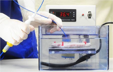
For that purpose, we constructed special wetlabs combining a basin containing isotonic saline solution with a thermostat and a pump to simulate an adjustable blood flow (Fig. 1). In these wetlabs, limited to in-vitro situations, ablation experiments can easily be performed.
Six identical workstations were equipped with this self designed wetlab simulating the blood`s temperature and flow and an own computer-controlled industrial RF ablation generator, allowing temperature- and power-controlled in-vitro RF ablations without or with open or closed cooling.
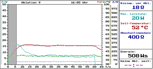
The software of the Osypka HAT 300s RF ablation generator allows an excellent didactic data analysis through continuous graphical and tabular recording and real-time-displaying of all relevant physical parameters, affecting the results of the in-vitro procedures (Fig. 2). Special screenshot and video capture software was installed on each workstation for easy documentation of parameters like radiofrequency power, temperature and impedance during the treatments. Any workstation’s screen can be displayed on additional large scale monitors for discussions of the observed effects in the training group.
Figure 1 Demonstration of in-vitro RF ablation on pork in the wetlab containing isotonic saline solution, heated to body temperature and simulating the cooling influence of the blood flow
By establishing special connection boxes, in-vitro experiments with catheters of different make and model can be performed. Thus, fundamental processes during RF ablation can be demonstrated and the special characteristics of different types of catheters and procedures can be revealed.
Figure 2 Demonstration of a dual-temperature-sensor in-vitro RF ablation. All relevant parameters are presented as diagrams: the courses of output power, the temperatures of both sensors and impedance. Furthermore, the applied energy as well as impedance before and after delivery of RF energy is displayed
Using this equipment the participants can learn, interactively, by measurements and visualization.
• differences between temperature and power controlled RF ablation
• differences in lesion size and geometry using standard 4 mm and 8 mm tip
• influence of the specific tip electrode angle to the myocardium on lesion size
• advantages and catheter handling using dual sensor technology
• differences between open1-4 and closed irrigated tip RF ablation
• influences of blood flow and cooling rate on lesion size
• effects of different tip materials on lesion size
• intracardiac electrogram recording
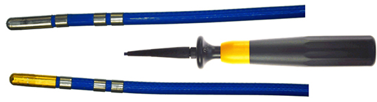
Figure 3 Identical Cerablate RF ablation catheters (Osypka AG) with 8 mm tip electrode of different materials. Top: standard platinum-iridium tip electrode; Bottom: novel massive-gold tip electrode; Middle: steering handle, identical for both catheters
During delivery of the radio frequency energy, the courses of power, one or two sensor temperatures and electrode impedance will be continuously displayed on each workstation monitor. It can by documented by screenshots and procedure protocol for every experiment. Furthermore, curve progressions can also be displayed on four large scale monitors for interactive discussions between professional teacher and the trainees.
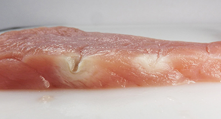
Figure 4 Comparison of lesions generated with 8 mm massive gold (left) and platinum-iridium electrode (right) using identical generator settings (T =65°C) and contact force
The training system was also used to investigate the influence of different catheter tip materials on lesion size. For example, we compared geometrical lesion size of identical catheters, differing in 8 mm massive gold and standard platinum-iridium tip electrode (Osypka AG) only (Fig. 3).
Figure 5 Plexiglass template, fixing a set of different diagnostic and therapeutic (ablation) catheters in the wetlab, in order to record artificial electrograms for test purposes
Compared to platinum-iridium, gold tip electrodes exhibit an almost four-fold thermal conductivity (3.17 versus 0.72 W/cm K). Thus, due to better cooling of the gold tip by blood flow, increasing RF power results in significantly larger and deeper lesion, as expected.5
Results of such experiments can also be used to reproduce in-vitro tests of new RF ablation catheters.
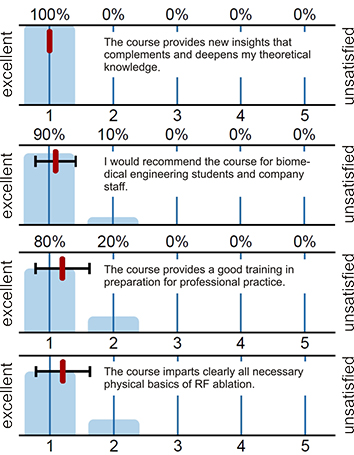
Figure 6 Excerpt from evaluation of the RF ablation hands-on teaching system
Results of these investigations, using identical RF ablation parameters, are shown in Table 1. Significant differences were found concerning lesion size by depth and diameter (Fig. 4). Using temperature-controlled RF ablation, the massive gold electrode tip showed an RF energy increase of up to 173% compared to platinum-iridium tip (Table 1). These results promise a reduction of ablation time, number of lesions and - consequently – decrease of fluoroscopy time.
The wetlabs can be equipped with a simplified plexiglass template to simulate different electrode arrays for electrophysiologic electrogram recording (Fig. 5). Connecting the output of a heart rhythm simulator to the wetlab, recording and particular filtering of intracardiac electrograms can de demonstrated by multi-channel EP-lab. Using this feature, the wetlab can also be used for tests and troubleshooting with diagnostic catheters, e.g. for elimination of noise.
During hands-on trainings for young electrophysiologists as well as for students of the biomedical engineering study path at Offenburg University of Applied Sciences, the in-vitro training system in physics of RF ablation provided excellent conditions to become acquainted with the theoretical and practical aspects of this method. This hands-on teaching system provides didactic simulations on the technical basics of the method. It allows a structured acquisition of practical knowledge. This ensures that physicians, company staff and biomedical engineering students receive a practical training, based on the needs of the clinical routine.
Table 1. Comparison between energy deliveries after 60 s RF ablations by platinum-iridium and massive gold electrode tips at different target temperatures using the Stockert SmartAblate G4 RF ablation generator (Stockert GmbH, Freiburg, Germany)
| Catheter tip material |
Energy (J) T = 45°C |
Energy (J) T = 55°C |
Energy (J) T = 65°C |
| Platinum-Iridium tip |
117 |
246 |
394 |
| Gold tip |
202 |
546 |
1075 |
| Difference (%) |
62 |
122 |
173 |
Evaluation of the didactic benefit was done by 10 of 12 trainees absolving the RF ablation physical basics course. On a scale between excellent (1) and unsatisfied (5), mean ranking was 1,2 of 10 different questions. Examples are shown in figure 6. Furthermore, this RF ablation teaching system was honoured for innovation in didactic of University education by a fellowship of the Association for the Promotion of Science and Humanities in Germany.
Finally the teaching system has proven to be a suitable environment for testing and evaluating of new catheters.


